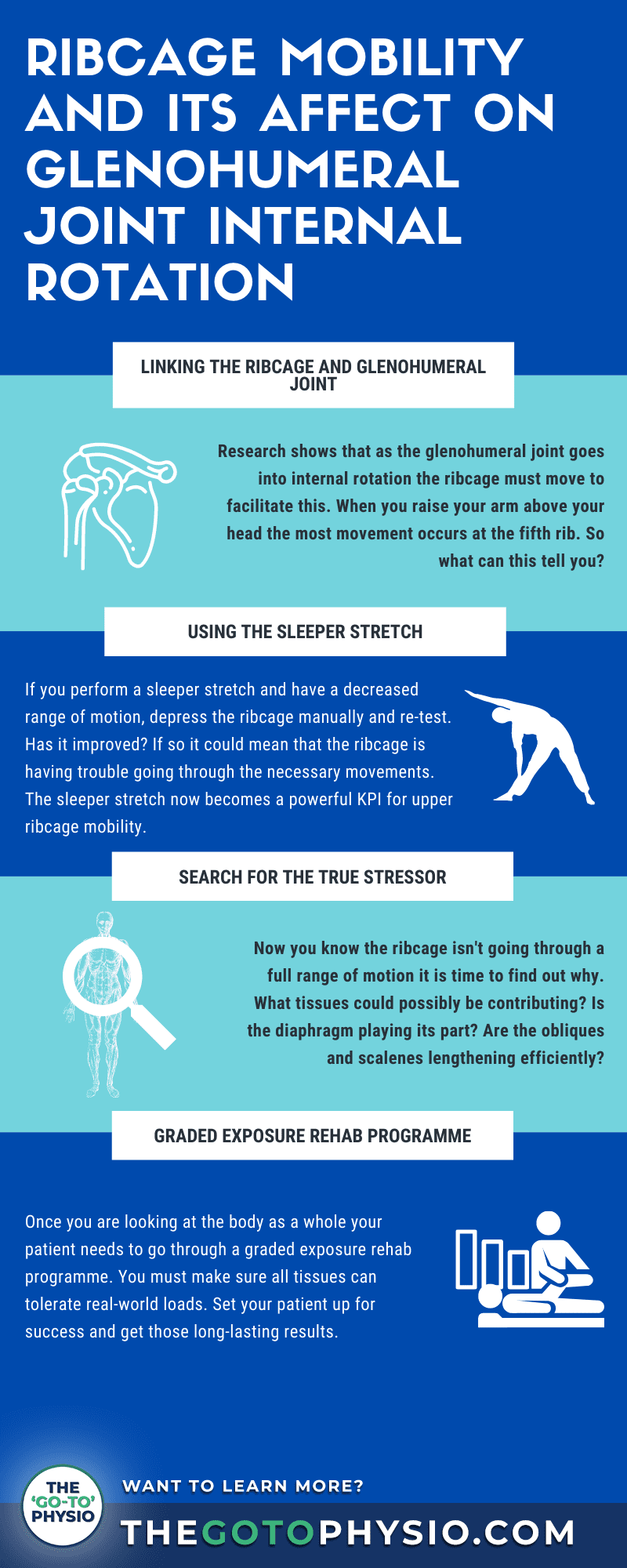
Every therapist has seen those tricky shoulder pain patients. You have tried everything you can but by the third or fourth session, they drop out. You are left wondering if you can help them at all. In this article, I want to share with you how patient adherence and long-lasting results may be closer than you think.
It is easy to get overwhelmed when looking at the whole body. When I had my first job in professional sports I felt that same feeling of being in over your head. But I quickly realised that if you don’t have the confidence and clarity to see the body in that way then you will only ever get short-term results
Today, I’ll show you a surprising link between two key players of the human anatomy that will shed light on those complex shoulder joint cases. You’ll learn how one simple approach can change the way you work for the better.
Read on to find out how the ribcage could hold the key to your success.
Anatomy Of The Glenohumeral Shoulder Joint
Before we begin to think about the human body as a whole it is important to note a few things about the glenohumeral joint.
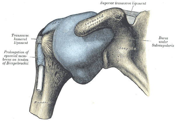
The glenohumeral joint or shoulder joint sits at the articulation between the humerus and scapula. In the groove between the humeral head and scapula lies the glenoid cavity, glenoid labrum, and articular cartilage.
Although this joint may seem simple there is a complex range of muscles and ligaments surrounding the joint capsule to ensure full shoulder range of motion. From putting on a t-shirt to throwing and catching, these muscles facilitate your patient’s real-world movement
The most commonly referenced tissues that surround the joint are the rotator cuff muscles. No doubt every therapist has seen a rotator cuff tear or rotator cuff dysfunction. This group of muscles is made up of the supraspinatus, infraspinatus, teres minor, and subscapularis.
These shoulder muscles are then further linked to the arm, working together with the deltoid muscle, biceps brachii, and others to facilitate full movement of the arm.
Understanding the anatomy of this joint is one thing but if a patient enters the clinic with a shoulder issue, how do you make sense of it? The complexity of the glenohumeral joint can be overwhelming. And being overwhelmed is a dangerous situation. But help may be closer than you think.
Using Glenohumeral Internal Rotation In Physiotherapy
Glenohumeral internal rotation is something I cover extensively in both the ‘Go-To’ Physio Mentorship and in my free diaphragm literature.
I was first introduced to the surprising benefits and key concepts of this motion by Ron Hruska of the Postural Research Institute (PRI). It is important to note that I don’t use a PRI approach but I have used the contents and structures of their courses, combined it with my own knowledge, and fed it into my own step-by-step system which I use in both professional sport and private practice and taught to over 600 therapists worldwide.
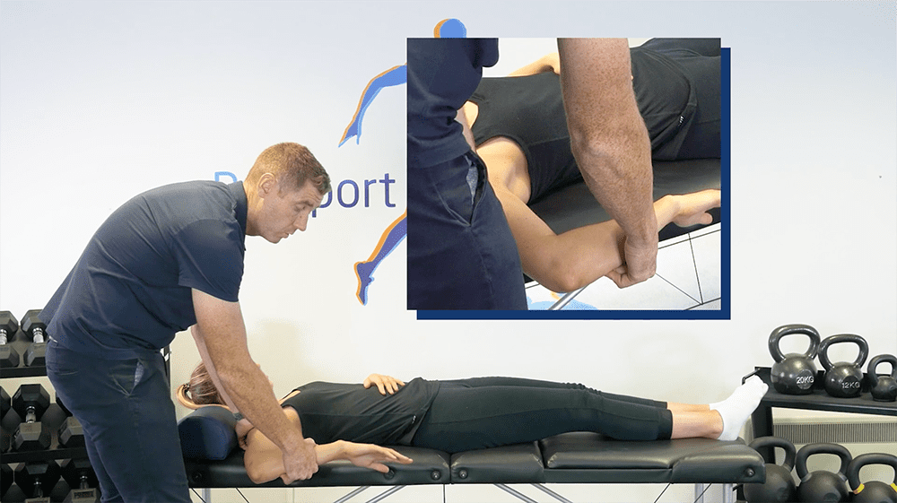
As a therapist, you may use this motion or others, such as the sleepers stretch test, to identify shoulder instability, impingement syndrome, or a rotator cuff tear but so many therapists do not understand the true power in this movement.
Very often in patients with joint and muscle issues, GH joint internal rotation range of motion will be decreased and there may be a pain experience.
Of course, if there is an issue with this exercise, then this would be something you would target with your graded exposure rehab plan, increasing load tolerance to improve rotation. But it can be overwhelming to know where to start.
To really understand what may be truly be going on with your shoulder pain patient you have to look at the bigger picture. Yes, you can worry about the glenohumeral joint capsule, the scapula, glenoid labrum, rotator cuff muscles, and various other ligaments and tissues, but unless you are asking these higher-level questions and looking at the body as a whole, then you are simply setting your patient up for failure.
One of the first places I look is the ribcage.
Why Does Ribcage Mobility Affect The Glenohumeral Shoulder Joint?
Using The Evidence
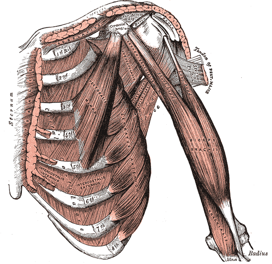
In a 2019 study, Tachihara and Hamada found that upper limb and ribcage movement are closely linked. Their findings suggest that when we elevate the upper limb most movement is found in the fifth, fourth and third ribs, decreasing toward the lower ribs.
Once you understand this, glenohumeral internal rotation becomes a useful key performance indicator for crossreferencing upper ribcage mobility. Not only does the ribcage influence the arm and upper limb motion but articles and research show ribcage mobility incurs large changes in lumbar curvature and pelvic movements.
Just take a wider view of what is really going on when you perform a sleeper test or test shoulder abduction, internal and external rotation. Once you are looking at the body as a whole and not just the joint, you’ll understand that for shoulder or glenohumeral internal rotation the ribcage must be mobile. Once the ribcage is mobilised this can then influence the lumbar spine and the thoracic spine.
All of these systems are closely interlinked. It isn’t just about the shoulder joint, the glenohumeral joints, the humeral head, or the joint capsule.
Pectoralis Major Stretch
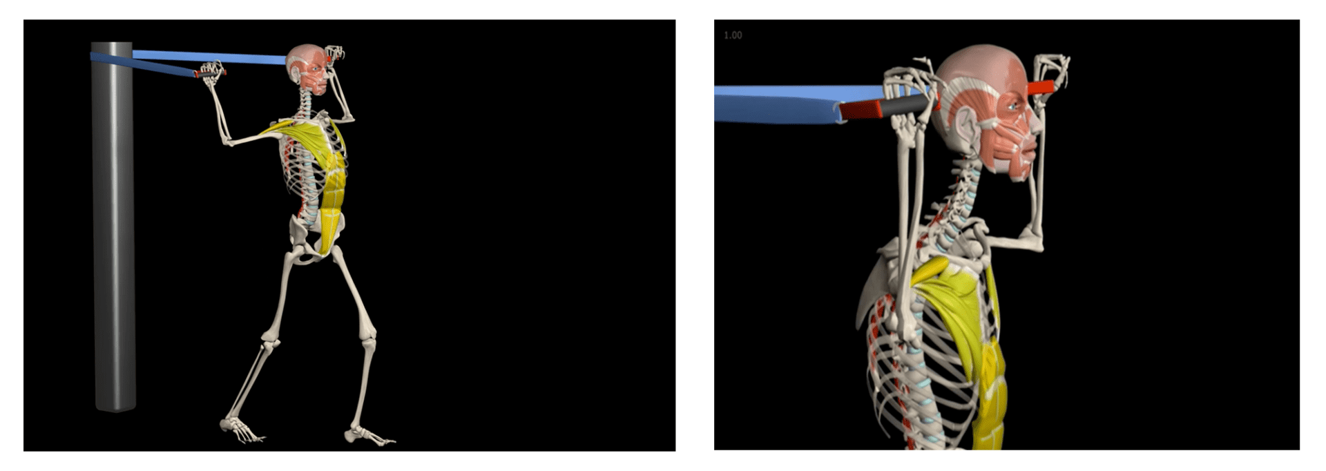
Take a patient doing the simple pectoralis major stretch as pictured below. What do you see? You may just see a pec stretch but I see an apprehension test.
If you take your thoughts off the pectoralis major muscle you can see the amount of motion in the ribcage as the subject goes through the motion. The ribcage, scapula, clavicle, rotator cuff, and many other tissues have to work to help the shoulder joint into this position.
In this position we aren’t just testing the glenoid labrum, a lot of other tissues have to work in order for this movement to be successful. If your patient doesn’t have the ability of the ribcage to protract and come forward then are they winding up the shoulder in the end range much earlier.
At first, we are getting depression and retraction of the ribcage. As the patient goes into glenohumeral external rotation the clavicular fibers of the pectoralis major stretch, along with the sternocostal fibers of the pectoralis major and anterior deltoid. There will also be protraction and elevation of the anterior ribs. In summary, external rotation at the glenohumeral joints couples with ribcage protraction and elevation, whereas internal rotation at the glenohumeral joints couples with ribcage depression and retraction.
So we can see that for the glenohumeral joint and shoulder to be successful your patient’s ribcage must be able to move effectively.
Why Wouldn’t The Ribcage Depress And How Could This Influence Internal Rotation?
Now, if you’re beginning to look at the body as a whole then you will start to realise just how many muscles, joints, and ligaments have to work together to allow everything to do its job efficiently.
We know that to achieve internal and external rotation of the glenohumeral joint we need the ribcage to depress, protract and retract. Once you have this you have the rotator cuff muscles, rotator cuff tendons, deltoid muscle, biceps brachii, and other tissues all working efficiently and providing stability.
If we can help the ribcage with free movement then we are setting the glenohumeral joint up for success when it comes to glenohumeral internal rotation. Allow for ribcage movement and glenohumeral joint rotation and you can set the whole body up for success.
To do this, you have to go back and look at the research. You have to ask yourself, what is attaching to the ribs that could influence the movements and cause an adaptation?
Here are a few more tissues that you will want to consider when it comes to free movement of the ribcage.
Serratus Anterior
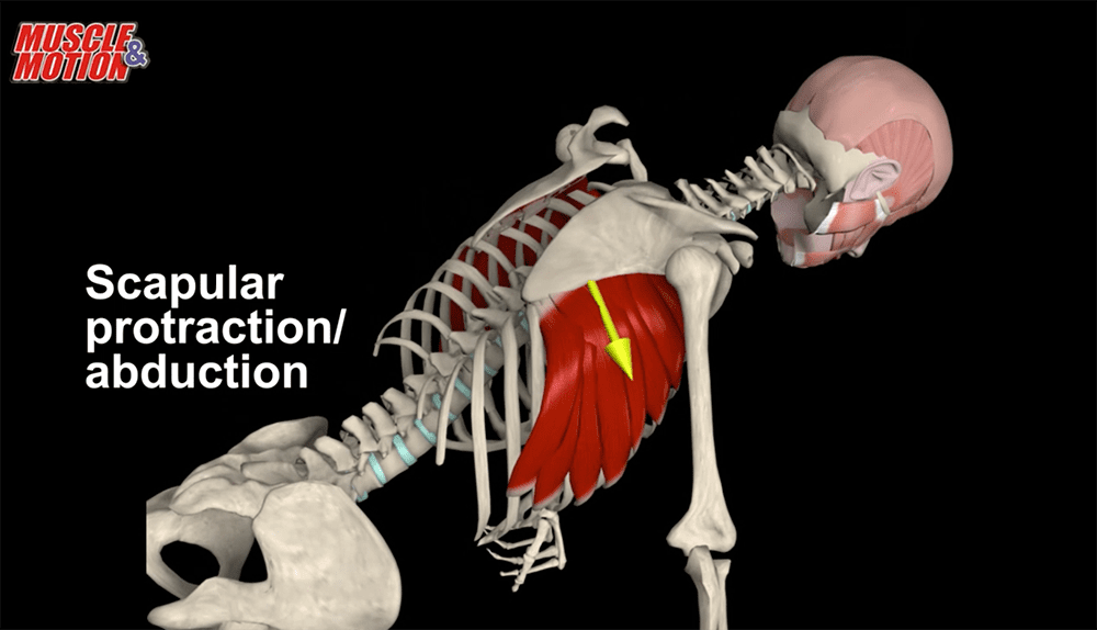
The serratus anterior muscles originate on the surface of the 1st to 8th ribs and attach to the deep aspect of the medial border of the scapula. It provides stability to the ribs and pulls the scapula forward allowing for anteversion and protraction of the arm.
If we consider a patient in the press-up position, then we can see just how much the ribcage protracts. The serratus anterior has to lengthen in order to facilitate a good position of the ribcage and allow successful glenohumeral internal rotation.
The Diaphragm
The diaphragm muscle is a dome-shaped structure situated at the bottom of the chest cavity, it inserts into the lower ribs and the lumbar vertebrae. It exerts control over the lungs for respiration keeping oxygen in the blood supply. When you inhale it flattens, causing a vacuum. When you exhale it lengthens.
The diaphragm plays a key role in allowing ribcage depression and motion. As the diaphragm lengthens and shortens to facilitate ribcage depression and retraction this then allows the glenohumeral articulation to enter internal rotation.
Beware… so many therapists believe the diaphragm is a magic bullet or a tissue that, when addressed will solve all of the shoulder joint or lumbar spine issues that walk through the door. Really it is only one piece of the puzzle. But you must understand, the diaphragm’s ability to go through a full range of motion has a serious impact on your patient’s ability to perform thoughtless and fearless movement.
Pectoralis Minor
The pectoralis minor originates in the third to fifth rib and attaches to the medial border and superior surface of the coracoid process of the scapula.
We know from the research that the fifth rib experiences the most movement when a patient reaches their arm over their head. In order to do this, the pectoralis major must play its part, lengthening and shortening when the ribcage goes into depression, protraction, and retraction.
If the pectoralis minor plays its part and lengthens on ribcage depression, then it can set the glenohumeral joint up for success with internal or external rotation.
Rectus Abdominis
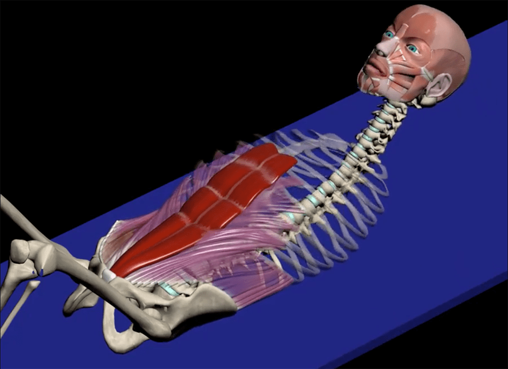
The rectus abdominis is a large structure tissue that provides stability to the trunk and allows for flexion of the thoracic and lumbar spine.
Like many other tissues we have mentioned, the rectus abdominis attaches around the fifth rib and so will have to lengthen, shorten and act as a stabiliser as the arm moves above the head and the glenohumeral enters into internal and external rotation.
Obliques
Much like the rectus abdominus the internal and external obliques support the core and provide trunk flexion and rotation.
The obliques also originate and attach to the fifth to the twelfth rib. Because of this close relationship with the fifth rib, as the arm and glenohumeral joint move, the obliques must be able to lengthen and shorten allowing the shoulder to go through the necessary motions.
Scalenes
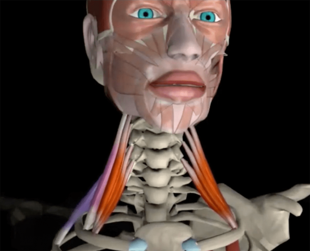
The scalenes consist of three pairs of muscles. The anterior, middle, and posterior scalenes. Their primary function is flexion of the neck but they also act as accessory muscles for respiration which is key in the context of ribcage movement.
The posterior scalene attaches to the second rib and the middle and anterior attach to the first rib. This means that when the ribcage depresses the scalenes must lengthen effectively.
If your patient is stressed and their breathing rate has increased then these accessory muscles will be working twice as hard. If the diaphragm does not have the ability to go through a full-motion and the accessory muscles are working harder, could this be contributing to the stiffness of the ribcage and in turn limiting glenohumeral joint internal and external rotation?
Next time you do have a shoulder pain patient, and manually depress their ribcage improving the internal rotation of the glenohumeral joint, consider hands-on treatment or stretches to target the protective tone in the scalenes rather than relying on breathing exercises alone.
By allowing these scalenes to lengthen distally, we allow depression of the ribcage and ultimately improve the internal rotation of the glenohumeral joint.
Every Tissue Working Together
I hope you have begun to see how just because your shoulder pain patient has a rotation restriction of the glenohumeral joint does not mean you should dive in with hands-on treatment trying to treat the joint, ligament, or anterior capsule.
The ability of the ribcage to depress has a close link to the ability of the glenohumeral joint to effectively rotate, but as we have seen there are far more tissues at play. All muscles must do their job effectively to set your patient up for real-world success.
Ultimately, your job as a therapist is to restore full range of motion. When presented with a knee joint injury with restricted flexion and extension, what do you do? Well, first of all you would probably turn your attention toward the joint capsule and some of those muscles that have an effect on the area when it is in motion.
This is exactly the same as what we are doing here with the ribcage. We are helping it go through a full range of motion as part of the treatment process to improve shoulder and glenohumeral joint movement.
The Effects Of The Respiratory System On The Glenohumeral Joint
Because the ribcage is so crucial to a shoulder condition and glenohumeral joint movement you must consider the respiratory system.
Now, I have already mentioned the respiratory system and the diaphragm and I must stress that this is no magic bullet. It is simply one part of your patient’s story. However, you must consider the interaction between the respiratory system, neurology, and the musculoskeletal system.
Prolonging the exhalation and restoring the ability of nasal breathing can have a widespread positive impact, lengthening the diaphragm, decreasing the protective tone of accessory breathing muscles such as the scalenes, and facilitating the patient’s ability to get back into rest and digest or their parasympathetic nervous system.
Of course, it isn’t always as easy as using three sets of ten deep breaths. What happens if your patient uses diaphragmatic breathing but they still have a protective tone around the scalene? You will only ever get those short-term changes.
Similarly, if you are lengthening the diaphragm effectively with your patient but they go back into the real world, where they are stressed and you haven’t these factors then you won’t get the long-lasting effects your patient needs and you deserve.
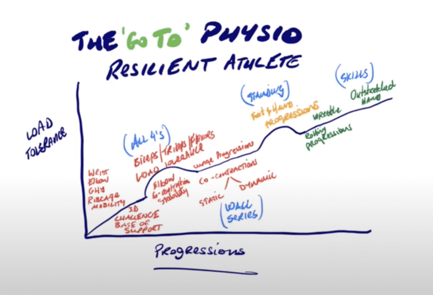
At this point is where you see the importance of having a rock-solid graded exposure treatment plan built around the person rather than the injury.
Using The 80/20 Rule In Your Clinical Practice
One of the most important things I teach in the ‘Go-To’ physio mentorship is the use of the 80/20 rule. It is an incredibly powerful tool and one that when used correctly will help you get those long-lasting results.
In the case of your shoulder pain and glenohumeral joint patient, it isn’t just about getting the scalenes or other contributing tissues to lengthen in order to facilitate shoulder function and internal rotation of the glenohumeral joint. That would be your 20%.
It is about finding the true cause and stressor specific to your patient that are causing the scalenes or the pectoralis major to have some kind of guarding which in turn prevents the ribcage from going through a full range of motion and the glenohumeral joint to effectively internally rotate. This is what you should be spending 80% of your time focusing on.
If we can get the ribcage to depress then we can set the glenohumeral joint up for success in internal rotation but finding the true cause as to why the ribcage won’t go through the motions is where the magic lies.
Building Patient Resilience Is The Key To Success
You have restored your patient’s ability to nasal breathe and you may have taken some protective tone away from the scalenes. Through this, you may have seen results in getting the ribcage to protract and depress and you have improved the loss of range and rotation in the glenohumeral joint. This may look like success but you need to remember this is the very first stage.
In real life, whether it is in professional sports or a non-sporting private practice patient you need them to be able to maintain the gains you achieve in the clinic in environments that might be physically or emotionally stressful.
This is where your graded exposure rehab plan comes into play. You must bridge that gap between lower and higher-level rehab and set your patient up for success in the real world.
Final Thoughts
Understanding the relationships between any number of key tissues is vital to getting the long-lasting results your patients need, whether they are a professional athlete or a non-sporting patient.
If you aren’t already, you must be asking yourself higher-level questions and beginning to clinically reason as to what the true stressor may be.
The next time you are assessing a shoulder internal or external rotation, or suspect a rotator cuff issue, do not get overwhelmed. Come back to these key points;
Anatomy Of The Glenohumeral Shoulder Joint
Do not underestimate the structure of the shoulders. From the glenoid capsule to the humeral head and rotator cuff muscles, there are far more components and soft tissue structures to think about than a simple articulation between the humerus and scapula. These tissues are never isolated.
The Relationship Between the Glenohumeral Joint And The Ribcage
If you know these elements are not isolated then you can begin to look at the whole body.
The articles and research I have shared tell us that both glenohumeral joint movement and ribcage movement are linked. Once you understand this relationship you can begin to look for the true cause of your patient’s issue.
Using The Sleeper Stretch To Measure Rotation
Once you know the movement of the glenohumeral joint and the ribcage are linked this can become a powerful key performance indicator.
In every session test and re-test your KPIs. If there is an improvement then you are on the right track. If there is a loss in range of motion in internal or external rotation then you may need to regress with your patient.
Why Won’t The Ribcage Depress and How Can This Help With Glenohumeral Joint Internal and External Rotation?
Remember there are so many contributing tissues that need to work efficiently to allow the ribcage to depress, retract and protract in order for the glenohumeral joint and shoulder to function efficiently.
Think about the diaphragm, other than exerting control over the lungs and keeping oxygen in the blood supply what effect does it have? Is it restricting ribcage movement and motion in the shoulders? Could the scalenes, obliques, or rectus abdominis be playing their part?
Use The 80/20 Rule
Use the ‘Go-To’ Physio 80/20 rule to divide up your time. In this case, spend 20% of helping the ribcage to depress and the shoulder to go into internal and external rotation and 80% of your focus on finding what the true stressor may be.
Building Patient Resilience
Just because you have restored the nasal breathing, helped the ribcage to move freely, and seen improvements in shoulder rotation does not mean you and your patient is in the clear.
This is just the very first stage. You must use a graded exposure treatment plan and set them up for success in the real world.
Get Long-Lasting Results
To get the long-lasting results your patient needs and you deserve you must look at the whole body. In the ‘Go-To’ Physio Mentorship I teach a structured step-by-step approach to your work. This means no more getting overwhelmed with complex shoulder pain cases, no more patient slump after session four. Just the confidence and clarity to help people in pain.
If you want more help gaining the confidence and clarity to achieve real results even with the most complex of shoulder pain patients, click the link below, book a FREE 30-minute patient adherence and rehab progressions strategy call and we’ll share with you the exact process we use for success with every patient.
